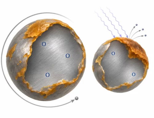
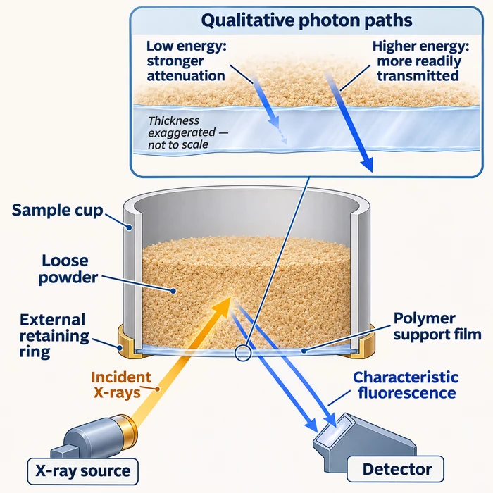
The support film lies in the fluorescence path. Its transmission depends on photon energy, composition, and path thickness; the schematic shows no measured transmission values.
A laboratory changes the support film on its loose-powder XRF cups, then sees a shift in reported composition. The retained powder has not changed, but its measurement path has. Before adjusting production settings, separate the film’s effect on detected fluorescence from the software’s conversion of that signal into concentrations.
Why the film changes the signal
On instruments that measure through the cup film, both the incident X-ray beam and the emitted fluorescence can be attenuated where their paths cross the film. Transmission depends on photon energy, film composition, and the mass of film along the path; NIST’s exponential attenuation relationship describes how, at fixed energy, material, and geometry, greater film thickness or an additional layer reduces transmission.
Low-energy fluorescence, such as Na Kα and Mg Kα, is particularly vulnerable. Polymer composition matters alongside thickness: at comparable thickness, polyester can attenuate these lines more strongly than polypropylene because their composition and density differ. Rigaku illustrates film-dependent transmission in its liquid-sample guidance; the same attenuation mechanism applies to fluorescence crossing a powder-cup film.
Why concentrations may behave differently
An intensity loss is not an equal percentage loss in reported concentration. Calibration converts net fluorescence intensities into concentrations, with matrix corrections and other processing between them. Check whether calibration specimens used the same film configuration and whether the method represents film absorption. Moriyama and Morikawa identify this correction as essential for Rigaku’s SQX fundamental-parameters analysis (section 3.2.2).
Also inspect the reporting basis. If one component’s unnormalized concentration is underestimated by film attenuation while the others remain unchanged, normalization to 100% can make those other components appear higher even though their true concentrations have not changed. Compare net intensities, unnormalized concentrations where available, and the final normalized report. The broader composition-testing framework helps define which constituent, preparation, and reporting basis the comparison must preserve.
Separate attenuation from other failures
Film additives can contribute elemental signals independently of absorption. Chemplex lists potential impurities by film type; Rigaku recommends checking each lot with a blank. Use the instrument manufacturer’s prescribed film-blank configuration, including backing material where required, rather than assuming an empty cup is adequate. Inspect cups for pinholes, stretching, wrinkles, poor seating, and sample loss. Leakage can contaminate the instrument.
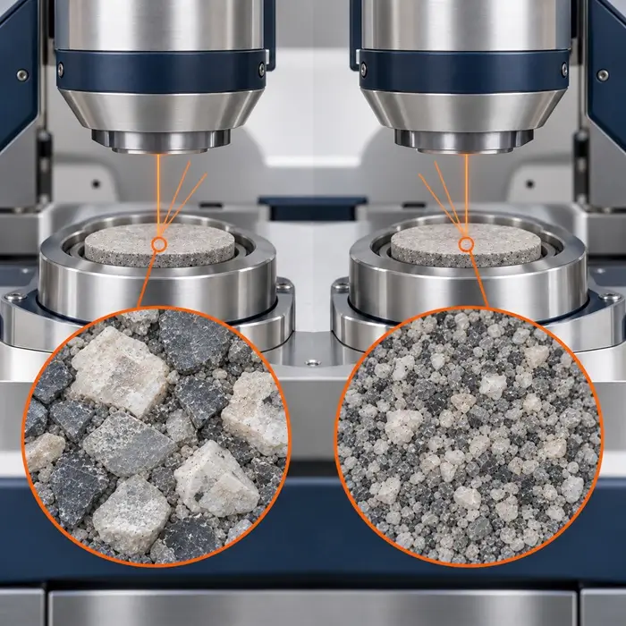
Keep powder depth and presentation consistent. At effective infinite thickness, additional specimen mass no longer materially changes the relevant fluorescence intensity. Below that condition, reduced intensity can reflect less contributing material rather than a composition change. Check this by comparing results as specimen mass per exposed area increases, holding preparation and geometry fixed; no single thickness or mass applies to every analyte and matrix. Preserve particle preparation too: grain-size and mineral-distribution effects provide additional reasons why nominally identical powders can give different XRF responses.
Run a controlled film comparison
- Take representative portions of one well-homogenized retained powder. Prepare independent cups for each film configuration. Record film product, polymer, nominal thickness, lot, layer count, and condition.
- Hold cup geometry, powder preparation, filling and packing procedure, measurement settings, and atmosphere fixed. First compare net intensities under identical acquisition conditions. Then evaluate reported concentrations using the appropriate film settings and validated corrections for each configuration.
- Measure appropriate film blanks and a certified reference material suited to the matrix, analytes, and concentration range under both configurations. Compare spectra or net intensities alongside concentrations and reference recoveries.
A repeatable film-dependent intensity shift supports a film-related explanation; acceptable reference recovery tests whether the method handles it adequately. If recovery deteriorates, resolve film settings, calibration compatibility, or preparation faults before adopting the new film. Document the validated configuration and its acceptance criteria in the laboratory method.
Technical Basis / Sources
- Moriyama, Takao, and Atsushi Morikawa. 2017. “Sample preparation for X-ray fluorescence analysis VII. Liquid sample.” Rigaku Journal 33(1): 24-29.
- Brouwer, Peter. 2018. Theory of XRF: Getting acquainted with the principles. 5th revised edition. Malvern Panalytical. ISBN 90-9016758-7.
- Hubbell, J. H., and S. M. Seltzer. X-Ray Mass Attenuation Coefficients. NIST Standard Reference Database 126, NISTIR 5632. DOI: 10.18434/T4D01F.
- Chemplex Industries, Inc. “Thin-Film Technical Data.” n.d.
- Takahashi, Gakuto. 2015. “Sample Preparation for X-ray Fluorescence Analysis III. Pressed and Loose Powder Methods.” Rigaku Journal 31(1): 26–30.