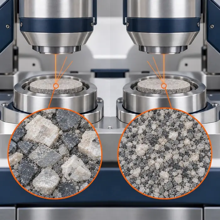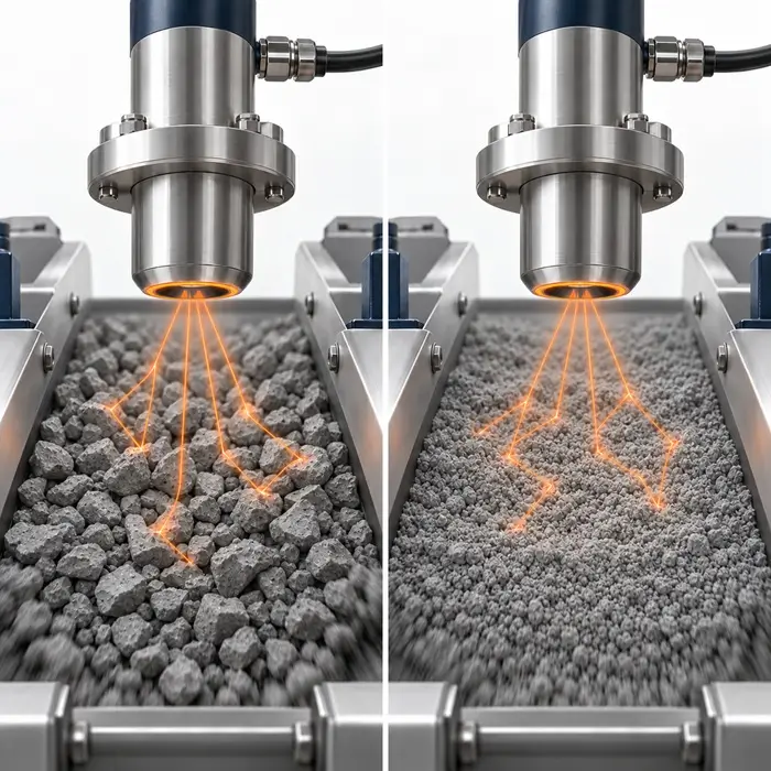
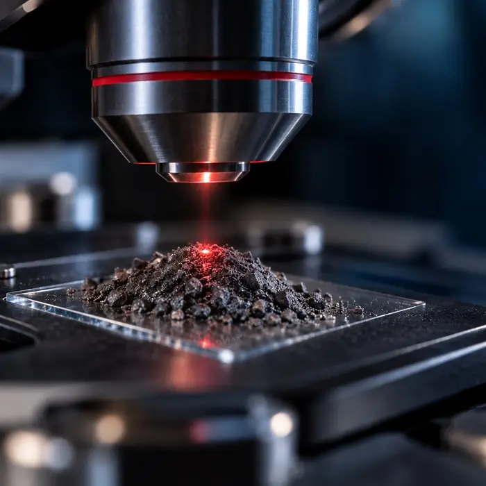
Two measurements of the same powder show different peak positions and intensities after an analyst increases laser power. Is the difference evidence of a material change, or did the measurement alter the specimen?
Why Can a Stronger Signal Become Misleading?
Absorbed laser energy causes localized heating. Temperature changes can shift vibrational frequencies, broaden bands, or modify relative intensities. Severe exposure can permanently transform or damage material.
Risk depends on excitation wavelength, absorption, focused spot size, thermal contact, and exposure duration.
Dark or colored powders often need greater caution than weakly absorbing pale powders. Metallic powders are not inherently safe: their reflectivity varies with wavelength, and poorly connected fine particles may still heat strongly. Thermally sensitive pharmaceutical powders also require low-power checks. No universal safe setting exists.
What Did the 2026 Study Demonstrate?
Li et al. (2026) examined loosely deposited aerosol-particle layers of chromium(III) oxide (Cr₂O₃), multiwall carbon nanotubes, and crystalline silicon, rather than bulk powders consolidated in sample cups.
They tested laser powers of 50–500 mW, integration times of 1–60 seconds, and deposit thicknesses of 0.05 and 0.5 mm.
Thicker chromium oxide and nanotube deposits developed ablation pits, while thinner layers retained recognizable bands even at 500 mW.
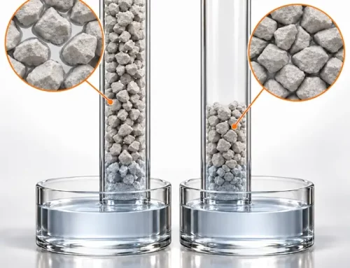
A likely explanation is that thin layers transfer heat more effectively to the supporting substrate, while thicker porous deposits restrict heat transfer from upper particles.
Controlled heating from 30 to 450°C reduced Raman band intensities, and the silicon band shifted to lower wavenumbers.
COMSOL simulations showed temperature rising within 800 milliseconds. Heating on this timescale helps explain why laser power dominated damage risk and why shortening a multisecond acquisition does not reliably prevent thermal artifacts.
Four Checks Before Assigning a Material Change
Check 1: Establish Power Dependence
Increase laser power stepwise, acquiring each spectrum at a fresh, comparable sample location. Keep wavelength, objective, focus, and acquisition time constant.
Compare peak positions, widths, relative intensities, and backgrounds.
If new bands appear, exclude measurement artifacts before assigning another phase. Thyr and Edvinsson (2023) describe false peaks caused by stray light, cosmic rays, optical components, and contamination.
Check 2: Quantify Heating and Check for Damage
Following each higher-power acquisition, remeasure its location at the initial low power and compare it with an untouched spot.
Recovered peak positions support reversible heating; persistent changes or visible pits suggest damage.
For crystalline silicon near room temperature, the Raman band around 520 cm⁻¹ shifts downward by approximately 0.02 cm⁻¹ per kelvin (Balkanski et al., 1983).
Other materials require their own calibrated temperature-sensitive bands. An adjacent silicon reference can check spectral stability, but cannot directly measure the temperature reached by a different powder particle.
For powders containing anatase TiO₂, the band near 143–144 cm⁻¹ offers a calibrated alternative (Zani et al., 2021).
Where signal strength permits, the anti-Stokes/Stokes intensity ratio provides another temperature estimate. Correct for detector spectral response; low-wavenumber bands give the strongest anti-Stokes signal and are usually the practical choice.
Check 3: Control Exposure and Sample Presentation
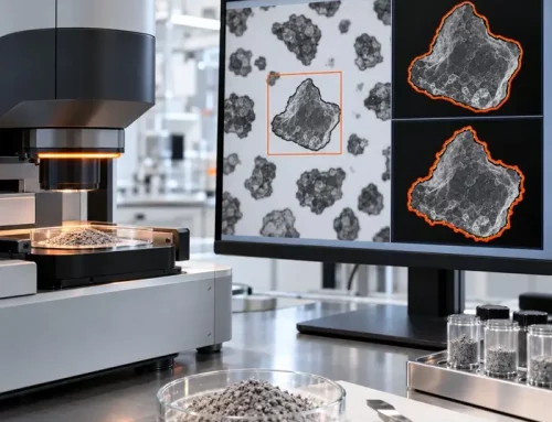
Record integration time, number of accumulations, deposit thickness, substrate, and powder distribution.
Compare equivalent preparations. A suitable heat-conducting substrate can improve heat dissipation, provided it does not interfere with the analysis.
Check 4: Verify Optical Conditions
Record excitation wavelength, microscope objective, approximate laser spot diameter, and power measured at the sample, rather than nominal laser output alone.
Foucher (2022) demonstrated that objective selection and beam geometry influence heating in material-dependent ways.
Reduce power or distribute acquisition across fresh sample locations when necessary.
Decision rule: Treat a Raman spectral difference as a material change only when it persists under validated, non-damaging measurement conditions.
Frequently Asked Questions
Technical Basis / Sources
- Li, J., Huang, J., Naz, S., Zhan, Y., and Zheng, L. (2026). Effect of measurement parameters and laser-induced heating on the Raman measurement of aerosol powders. Talanta, 301, 129259.
- Foucher, F. (2022). Influence of laser shape on thermal increase during micro-Raman spectroscopy analyses. Journal of Raman Spectroscopy, 53(3), 664–676.
- Thyr, J., and Edvinsson, T. (2023). Evading the Illusions: Identification of False Peaks in Micro-Raman Spectroscopy and Guidelines for Scientific Best Practice. Angewandte Chemie International Edition, 62(43), e202219047.
- Balkanski, M., Wallis, R. F., and Haro, E. (1983). Anharmonic effects in light scattering due to optical phonons in silicon. Physical Review B, 28(4), 1928–1934.
- Zani, V., Pedron, D., Pilot, R., and Signorini, R. (2021). Contactless Temperature Sensing at the Microscale Based on Titanium Dioxide Raman Thermometry. Biosensors, 11(4), 102.